Supporting documents
Clinical history and notes
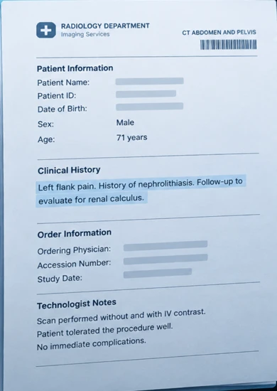
- Clinical history
- Requisition
- Technologist notes
- Scanned paperwork
What problem we solve, by care setting.
Smart Dictation® brings available study details together so you do less repetitive work. Review what is already prepared, dictate only the new or important findings, and save time on the report.
01Context prepared
02Your interpretation
03A draft to review
Clinical history and notes
Structured measurements
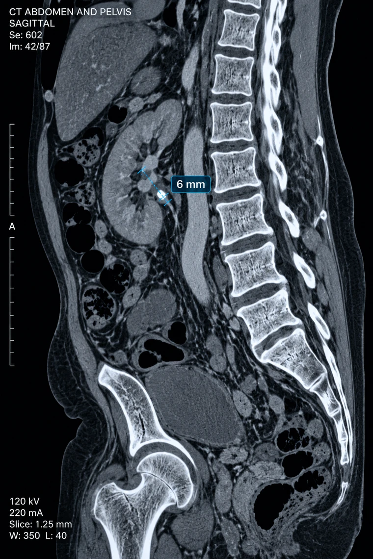
Appropriate reporting template
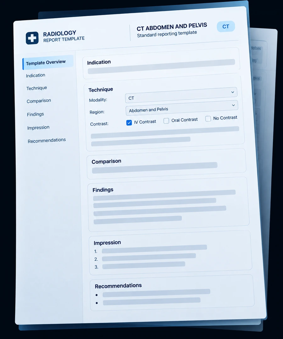
Check the prepared details and choose what belongs in the draft.
Available details prepared.Ready for your findings.
Review, correct and dictate
Illustrative content from synthetic supporting information.
Available history and prior-study details can populate your template. Measurements, notes and connected AI findings are inputs in Smart Dictation® for you to review and select for the draft.
Spend less time gathering, typing and dictating information already available.
Bring relevant clinical information into the read, without repeating it into the microphone.
Check the prepared details and add the findings that need your interpretation.
Shape AI-assisted reporting around the way you work. Set reporting preferences that help RADIN organize and present your findings in your preferred style.
Lung nodule right upper lobe increased in size, now 9 mm. Mild degenerative changes of the thoracic spine. New small right pleural effusion.
Known pulmonary nodule. Follow-up evaluation.
Prior chest CT.
New small right pleural effusion. Right upper lobe pulmonary nodule now measures 9 mm, previously 6 mm. Mild degenerative changes of the thoracic spine.
Illustrative customization with synthetic content. Every draft requires radiologist review.
Dictation AI and RADINA flag possible report discrepancies and help connect the next step.
Flag possible side, anatomy and measurement conflicts.
You decide what to change. Checks depend on configured modules and integrations.
Configured modules carry findings into communication, follow-up and operations.
Log calls and escalation.
Track recommendations to completion.
Extract codes and report RVUs.
Review discrepancies and addenda.
Assign studies by eligibility and priority.
Dictate key findings and review the draft before signing.
Standardize reports and track follow-up across the practice.
Connect reporting to your PACS and manage access centrally.
ONE PLATFORM
Worklist, viewer, reporting and AI in one place.
Explore RADIN PACS →YOUR EXISTING PACS
Use RADIN reporting with your viewer through configured integrations.
Explore RADIN Workflow →SEE IT WITH YOUR STUDY TYPES
Bring your study types and reporting preferences. We’ll show what Smart Dictation® prepares for you, and what is left for you to say.
AI-generated report content is reviewed, edited and signed by the responsible radiologist. All demonstrations use illustrative, synthetic information.